EPFL releases a free tissue atlas to help AI understand cancer
Life sciences
2 November 2026
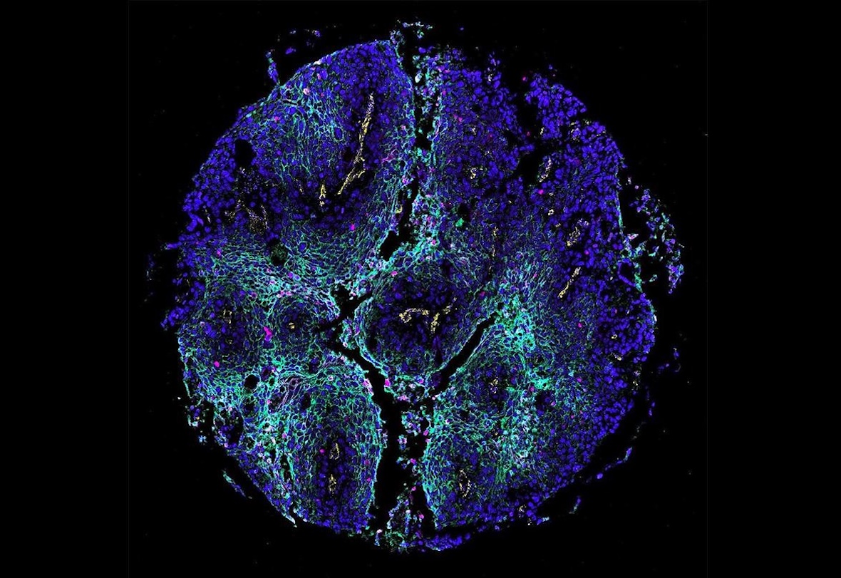 EPFL’s spora atlas gathers 13,546 images of tissue samples from 5,477 patients. | © EPFL/AIMM
Built with partners in Zurich and Geneva, spora is a spatial proteomics atlas that EPFL describes as the world’s largest freely available collection of its kind, designed to speed up AI research on how diseases develop.
EPFL researchers have released spora, a reference atlas of spatial proteomics images and medical data, together with the European Bioinformatics Institute (EMBL-EBI), which hosts it in its BioImage Archive. Spatial proteomics measures dozens of proteins while preserving their location in tissue, showing how cells are arranged and interact. EPFL describes spora as the world’s largest freely available collection of such images and data.
The atlas gathers 13,546 multiplex images from 5,477 patients, covering 14 tissue origins and five imaging technologies, with paired histopathology images where available. The team spent two years organizing the images, standardizing protein annotations, matching medical information and identifying individual cells, so that datasets from different labs, which often use different equipment, proteins and storage formats, can be combined more easily.
Tools for AI research
spora comes with three components: spora[data], with images and annotations in a common format, spora[io], a Python library to load tissue regions, combine cohorts and prepare training data, and spora[bench], a benchmarking suite with tasks such as identifying cells, classifying cell types and predicting clinical variables. Researchers can use it to test whether findings hold across studies, whether models recognize cell types across different protein panels and whether a model trained at one hospital works on images from another. Accompanying AI models were trained on large-scale computing through the Swiss AI Initiative.
The project was led by Charlotte Bunne, who heads the Artificial Intelligence in Molecular Medicine (AIMM) group at EPFL, with the University Hospital Zurich, the University of Zurich, the Geneva University Hospitals (HUG) and the University of Geneva, and was developed under the National AI Initiative for Precision Oncology. “Our goal is to turn what we learn across thousands of tissue samples into insights that can help individual patients,” says Bunne. The team plans to add more tissues, diseases and imaging technologies.