
HUG and UNIGE unveil the next frontier in Alzheimer’s diagnosis
Life sciences
15 August 2023
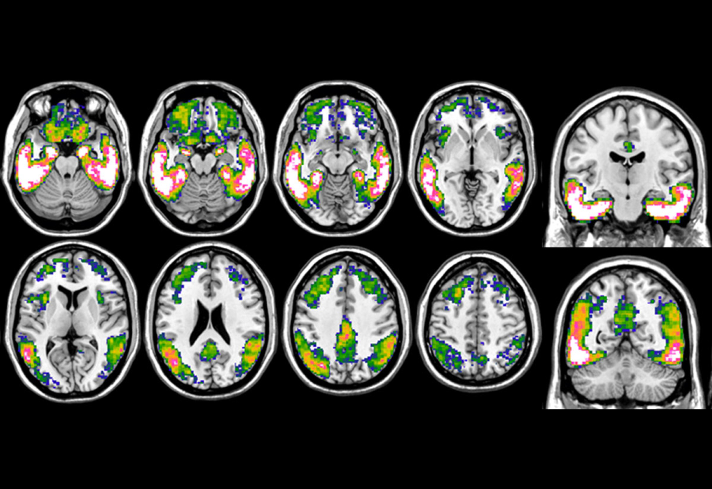 Tau imaging with 18F-Flortaucipir PET in Alzheimer’s disease. The figure shows a prototypical tau accumulation pattern, obtained by comparing tau load of patients with Alzheimer’s disease vs healthy controls. The blue-to-white color scale indicates more pronounced tau loads, with pink-white areas representing those with the highest accumulation. | © UNIGE
HUG and UNIGE’s tau PET imaging offers a transformative leap in Alzheimer’s diagnosis, promising early detection and brighter futures for millions affected.
The world of medical science is constantly evolving, with breakthroughs emerging that have the potential to transform our understanding of various diseases. Alzheimer’s, a notorious neurodegenerative disease known for its gradual memory erosion, has been a significant area of research focus. Recent advancements in tau PET imaging, developed at the University of Geneva (UNIGE) and the Geneva University Hospitals (HUG), promise to reshape the diagnosis and early intervention of this debilitating disease.
Alzheimer’s Disease is marked by the buildup of neurotoxic proteins in the brain, notably amyloid plaques and tau tangles. Understanding these accumulations is vital, especially considering the silent evolution of Alzheimer’s pathology over years, sometimes even decades. Early diagnosis can lead to timely interventions, which might slow down disease progression.
Currently, positron emission tomography (PET) plays a vital role in diagnosing Alzheimer’s. Valentina Garibotto, who spearheaded this research and holds prominent positions at both UNIGE and HUG, clarifies, “PET allows the visualization of specific pathological processes in the brain through the injection of low-level radioactive tracers.”
While tracers for amyloid and glucose metabolism have been in existence for a while, they do not provide a comprehensive insight into the complexities of Alzheimer’s. Herein lies the game-changer: Flortaucipir, an FDA-approved radiotracer developed in 2020, specifically targets the tau protein, enabling researchers to detect tau accumulation and understand its distribution and clinical implications in the brain.
The gold standard in predicting Alzheimer’s progression
In a study at the HUG Memory Centre with 90 participants, researchers found tau PET imaging to be the most predictive of Alzheimer’s-induced cognitive decline. A key contributor to the study, Cecilia Boccalini noted its accuracy even in patients with minor symptoms. While amyloid plaques indicate Alzheimer’s, it’s tau that aligns with clinical symptoms, signaling the disease’s progression.
The HUG and UNIGE teams emphasize the promise of early detection paired with new treatments. Understanding tau’s distribution in the brain is crucial for addressing Alzheimer’s complexities, and incorporating tau PET imaging into routine clinical evaluations can help map personalized therapeutic pathways for patients, bringing hope to millions affected by this disease.